Non-invasive longitudinal tracking of stem cells to advance cell therapy
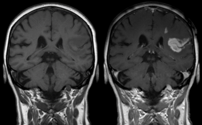
Jeffrey Struss Associate Scientist, Nanomaterials Magnetic Resonance Imaging (MRI) is one of the most commonly used non-invasive imaging techniques in medicine. Allowing the clinician to visualize the structure of the tissues inside the body in high resolution without the need for vivisection. With the dramatic enhancements to in vivo imaging and the growth of cellular-based therapeutics, the need to track cells as they travel, over time, through the body has continued to grow. Whether immune cells, stem cells, cancer cells or native cells, the ability to see how cells travel and congregate throughout the body can provide great insight into various issues such as cancer, auto-immune disorders, wound healing and aging. One of the ways to improve imaging in MRI applications is via the introduction of a contrast agent. Contrast agents interact with the magnetic fields of the MRI to enhance the appearance of internal biological structures. The image below shows a stroke patient before the administration of a contrast agent (left) and post administration of a contrast agent (right). The use of the contrast agent in the right image makes it very clear where the stroke occurred and the magnitude of tissue affected by the stroke.
The most commonly used class of MRI contrast agents are Gadolinium-based agents. Due to the highly toxic nature of ionic gadolinium, it cannot be injected directly. Instead, it is typically administered in a chelated format using chelation agents such as diethylenetriaminepentaacetic acid (DTPA) or tetraazacyclododecane tetraacetate (DOTA). Unfortunately, chelation of the gadolinium ion reduces its effectiveness as an MRI agent.
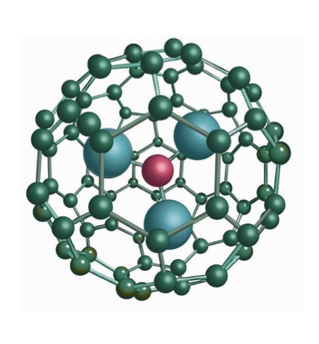
As a result, much work has been done to find more effective gadolinium-based preparations. Enter the Trimetasphere© from Luna Innovations. The Trimetasphere© nanostructure incorporates a Gd3N cluster encapsulated inside of a C80 fullerene cage. This proprietary and innovative structure allows for the direct administration of ionic gadolinium in a safe and effective manner, bypassing most of the major issues found with other ionic gadolinium preparations. By encapsulating the toxic gadolinium ions in a robust and biologically-compatible cage, it provides all of the benefits of ionic gadolinium without any of the drawbacks. Recently, MRI has been finding new use in the field of cellular therapy. Cellular therapy involves the therapeutic application of specific cells in order to treat disease. These cells can be everything from a variety of stem cells to genetically modified whole cells. Currently, the analysis of the migration of these applied cells can only be performed post mortem. As a result, there is a strong push to solve the challenge of tracking and analyzing the migration of these cells in vivo. MRI, coupled with contrast agents, has the capabilities to solve this problem. An important study was performed recently employing Luna’s novel Trimetasphere© agent, evaluating the safety and efficacy in longitudinally tracing stem cells in the lungs. In the first phase of this experiment, amniotic fluid stem (AFS) cells were incubated with varying concentrations of the Trimetasphere© reagent. The cells were evaluated both for their viability and their ability to proliferate. They were also evaluated, via PCR, for the activation of seven different pathways associated with cellular stress. For all of the concentrations studied, there were no apparent ill effects.
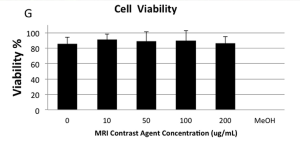
The figure above, shows the effect of varying concentrations of Trimetasphere© on both cell viability and absorbance. It is clear from the first graph that even at very high concentrations of Gd-Trimetasphere©, there is no additional cellular death when compared with the positive control. In the next step of the study, a mouse model of a simulated lung injury was generated via targeted radiation exposure. Trimetasphere© tagged stem cells were administered via tracheal intubation to the mouse and were observed as they travelled through the lungs.
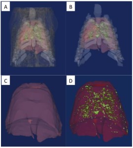
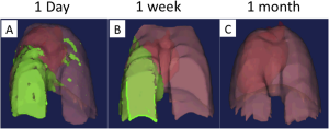
These images clearly illustrate where the Trimetasphere© labeled stem cells have localized in the lungs. Much of the localization is on the side with the induced injury, as expected. Initially, some broad distribution to both sides of the lungs occurs, but at the time of 1 week all of the remaining cells are localized to the injured side. By one month, no tagged stem cells can be detected. This complete clearance is also important for longitudinal studies for tracking additional applications and results. It is clear that Trimetasphere© contrast agents provide a safe, accurate and reliable method for the tracking of stem cells post-application in vivo. The novel and ground-breaking technology allows researchers to safely and accurately track the progress of cells throughout the body in real time, without the necessity of post mortem analysis. As a result Trimetasphere© contrast reagent allows for more rapid turnaround of experiments with faster acquisition of data and more detailed longitudinal information. With this data, researchers can more accurately generate conclusions and design further experiments. Additionally, due to the ability to analyze in vivo, multiple experiments can be performed on a single subject reducing the run-to-run variability.